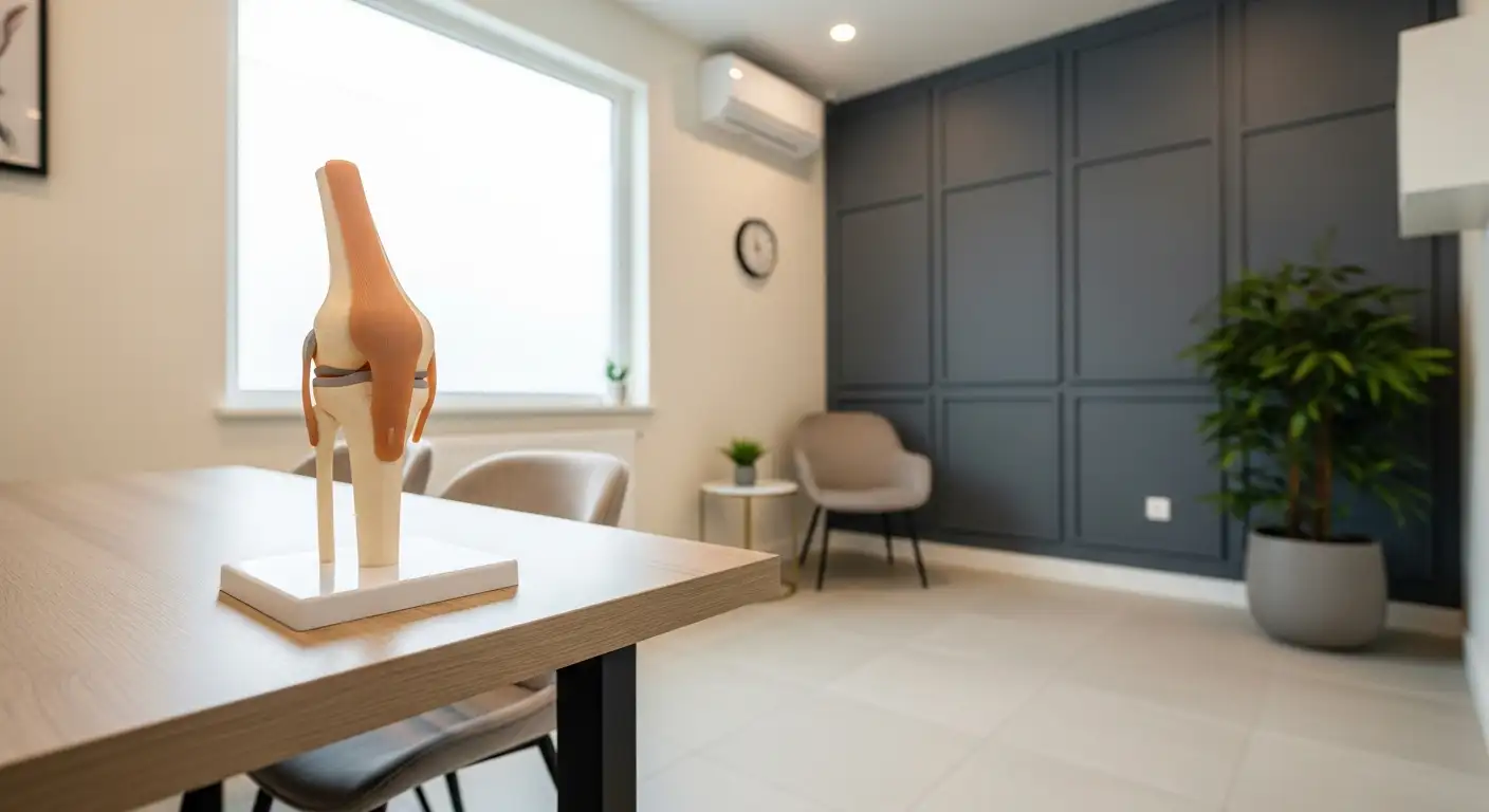
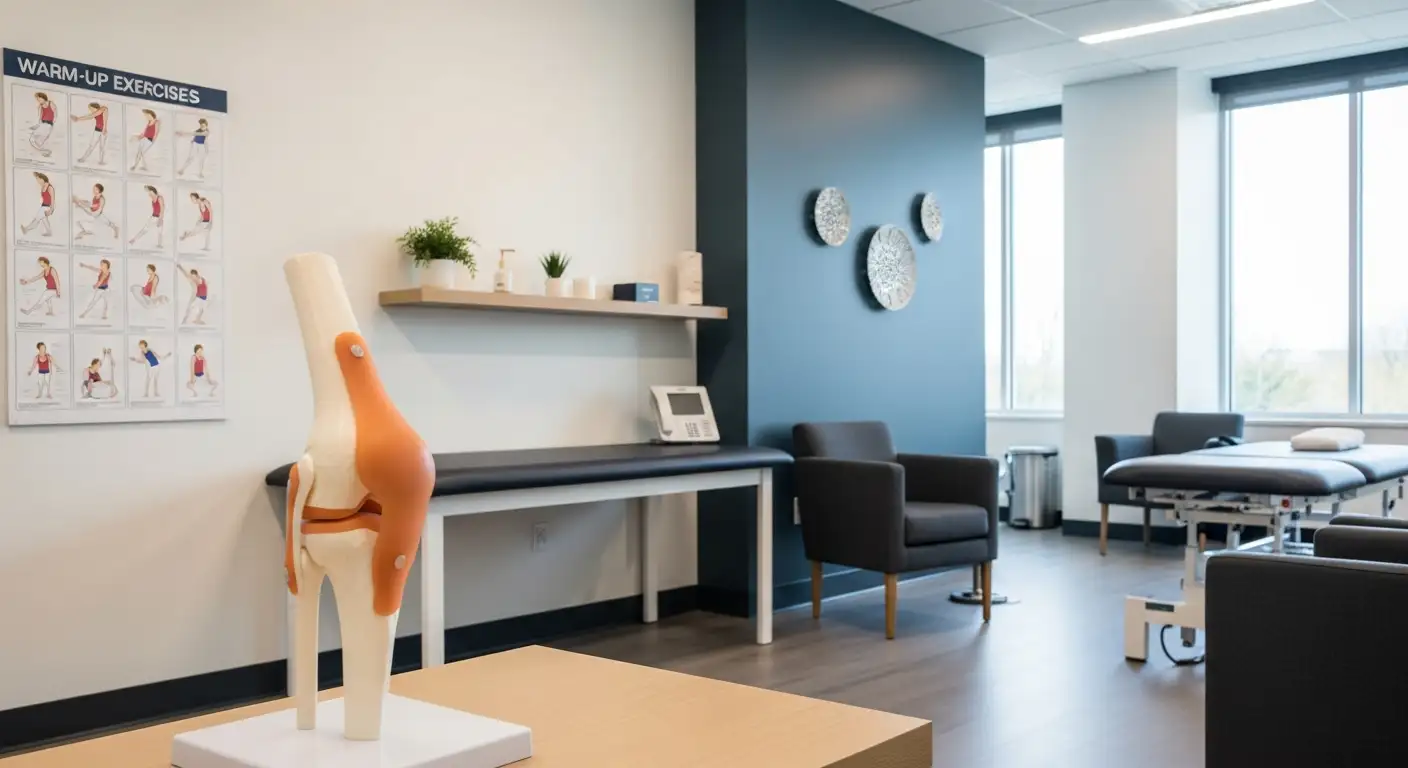
Understanding Knee Pain
Knee pain can stem from various conditions, with chondromalacia patella being one of the prominent causes. Understanding the causes and symptoms associated with knee pain is crucial for anyone experiencing discomfort or seeking insight into their condition.
Causes of Knee Pain
Chondromalacia patellae, often referred to as "runner's knee," arises from the softening of the cartilage on the kneecap. This condition is common among young athletes, but older adults with knee arthritis may also experience it. Factors contributing to chondromalacia include:
- Age: Adolescents and young adults are at a higher risk due to rapid muscle and bone development.
- Sex: Females are more predisposed to developing runner's knee.
- Foot Structure: Individuals with flat feet may be more susceptible.
- Previous Injuries: History of knee injuries can increase risk.
- Activity Levels: High activity levels can contribute to the condition.
- Arthritis: Existing knee arthritis may also play a role in the development of chondromalacia.
These factors can lead to the softening, tearing, or erosion of the hyaline cartilage at the back of the kneecap. For further information, visit healthline.
| Risk Factor | Description |
|---|---|
| Age | Adolescents and young adults at high risk |
| Sex | Females more likely to develop the condition |
| Foot Structure | Flat feet may increase susceptibility |
| Previous Injuries | History of knee injuries heightens risk |
| Activity Levels | High levels of physical activity impact risk |
| Arthritis | Existing knee arthritis contributes to chondromalacia |
Symptoms of Knee Pain
Those suffering from chondromalacia patellae often notice several symptoms that can complicate daily activities. Common symptoms include:
- Knee Pain: Often localized around the kneecap.
- Grinding Sensations: A feeling of grinding or crunching, particularly during movement.
- Increased Pain After Sitting: Pain may worsen after prolonged sitting or after activities that apply pressure to the knees, such as standing or exercising.
- Effusion: Swelling in the knee joint can occur.
- Wasting of the Quadriceps: Muscle atrophy may be observed in the quadriceps.
- Retropatellar Crepitus: A popping or crackling sensation behind the kneecap.
These symptoms may indicate chondromalacia, but they are not exclusive to this condition. A reliable diagnosis requires the exclusion of other potential causes of anterior knee pain. For more insights on symptoms and conditions related to knee pain, refer to NCBI.
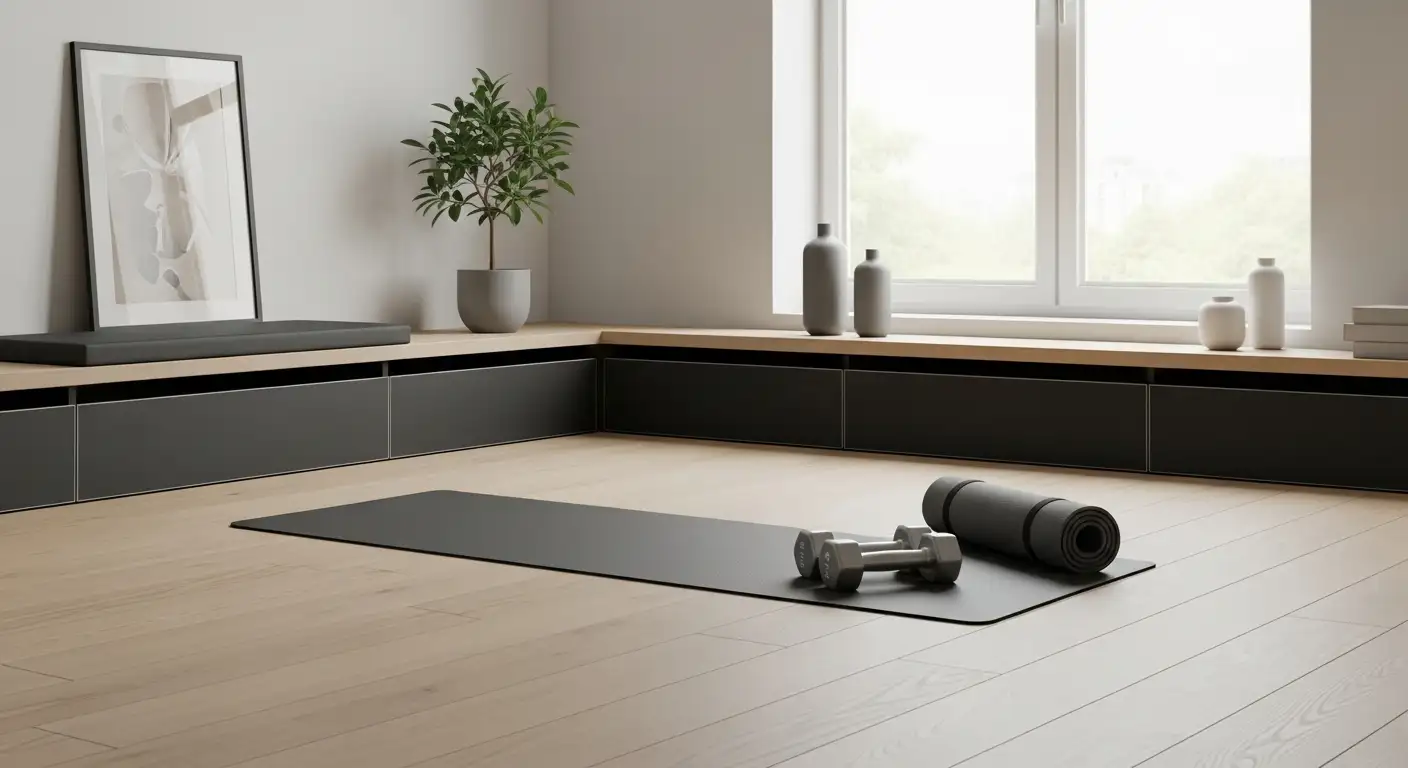
Non-Surgical Treatment Options
For individuals experiencing knee pain related to chondromalacia patella, non-surgical treatment options are often the first line of defense. These options include rest and ice therapy, physical therapy, and medications aimed at relieving pain and inflammation.
Rest and Ice Therapy
Rest and ice therapy serve as foundational treatments for managing knee pain. This approach helps reduce inflammation and allows the knee joint time to heal. Rest involves avoiding activities that put strain on the knee, particularly those that exacerbate the pain. Ice therapy should be applied for 15-20 minutes every few hours to decrease swelling and numb the pain.
| Treatment | Duration | Frequency |
|---|---|---|
| Ice Application | 15-20 minutes | Every few hours |
| Rest | As needed | Varies |
Consultation with a healthcare provider is essential to create a personalized plan that incorporates these techniques effectively.
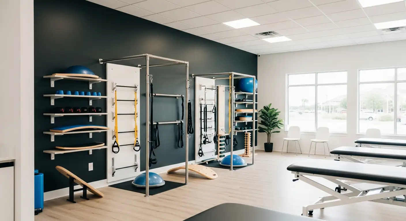
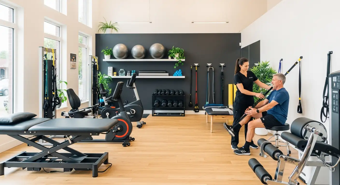
Physical Therapy
Physical therapy is crucial in managing chondromalacia patella and often focuses on strengthening the quadriceps and other supporting muscles around the knee. A physical therapist may develop a program that includes non-weight-bearing exercises and stretches such as band stretches or a gluteus maximus stretch. The following table illustrates potential exercises often recommended:
| Exercise Type | Goal | Example |
|---|---|---|
| Strengthening | Improve muscle stability | Quadriceps strengthening exercises |
| Flexibility | Increase joint range of motion | Stretching exercises |
| Low-impact | Reduce stress on the knee | Swimming or cycling |
Patients should work with a health professional to ensure safe execution of exercises geared toward knee rehabilitation.
Medications for Knee Pain
Nonsteroidal anti-inflammatory drugs (NSAIDs) are commonly used to alleviate pain and reduce inflammation in cases of chondromalacia patella. Medications such as ibuprofen may be suggested as part of the management plan. However, patients should consult with a healthcare provider to determine the most suitable medication and dosage for their specific needs.
In some cases, other treatment modalities such as autologous chondrocyte implantation or injection of mesenchymal stem cells might be considered beneficial, particularly if conservative measures do not yield satisfactory results [1].
For a comprehensive approach to knee pain treatment, it is essential to collaborate with an interprofessional team, including orthopedic surgeons and physical therapists, to devise a tailored plan that meets the patient's needs.
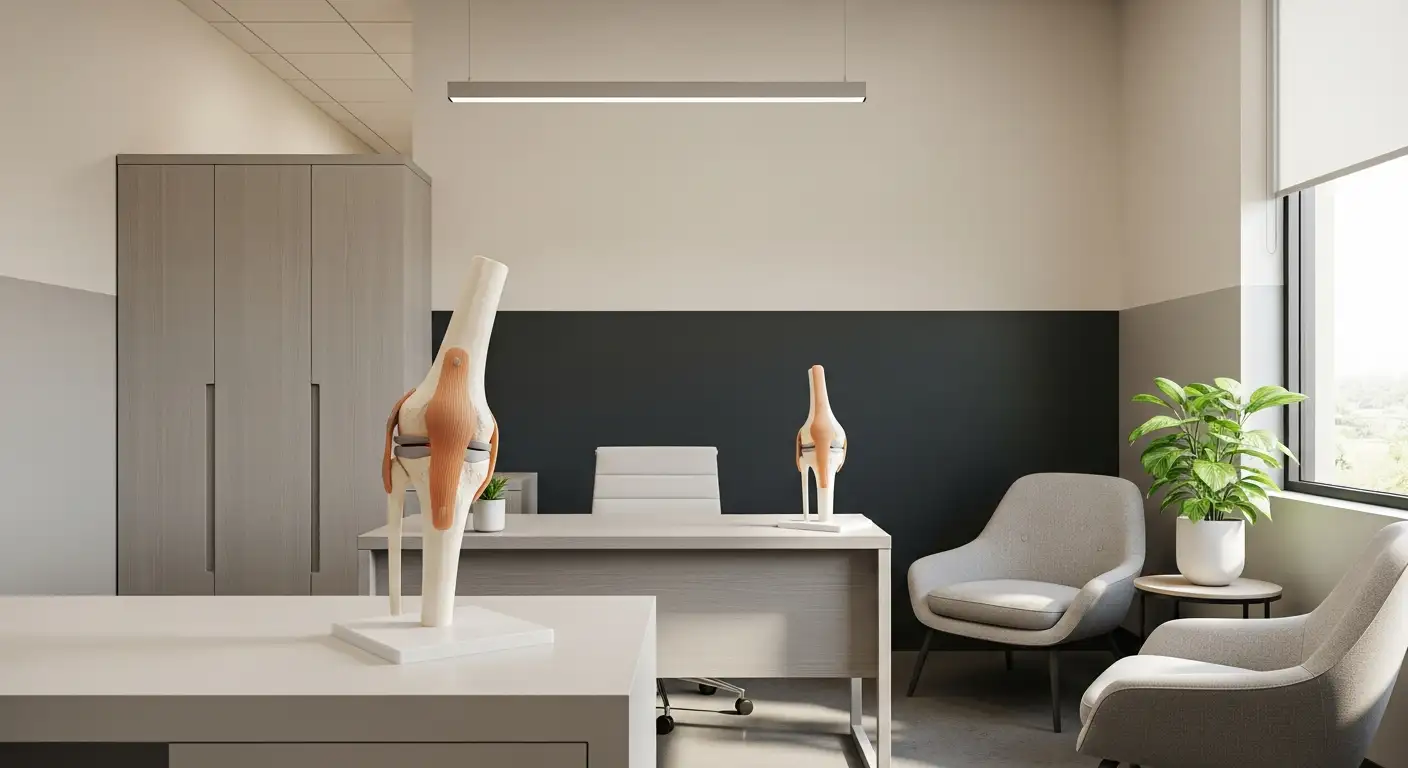
Surgical Considerations for Chondromalacia
When non-surgical options have not relieved knee pain from chondromalacia patella, surgical interventions may be considered. Two common procedures include arthroscopic examination and lateral release surgery.
Arthroscopic Examination
Arthroscopic examination is a minimally invasive procedure used to diagnose and treat knee joint issues, including chondromalacia patella. The surgeon makes small incisions around the knee and inserts a slender instrument with a camera, known as an arthroscope. This allows the physician to view the inside of the knee joint on a monitor and assess the extent of cartilage damage.
This procedure is typically performed as day surgery, meaning patients can return home on the same day. Depending on the severity of the damage, the surgeon may also treat any identified issues during the examination. The use of arthroscopy generally results in less postoperative pain and a quicker recovery compared to open knee surgery, which may require a longer hospital stay.
| Feature | Arthroscopic Examination |
|---|---|
| Procedure Type | Minimally invasive |
| Hospital Stay | Typically none (day surgery) |
| Recovery Time | Shorter recovery compared to open surgery |
Lateral Release Surgery
Lateral release surgery is another option for addressing issues related to chondromalacia. This procedure focuses on realigning the kneecap (patella) by releasing tight lateral structures, often referred to as the lateral retinaculum. This can help alleviate pressure on the cartilage and reduce pain.
Lateral release can be performed arthroscopically or through an open surgical approach, depending on individual circumstances. The decision on which method to use often depends on the severity of the cartilage damage and the patient's overall knee alignment. Patients undergoing this procedure are typically advised on postoperative care, such as using crutches for mobility and wearing a knee brace to stabilize the joint.
Summary of Surgical Options
In summary, both arthroscopic examination and lateral release surgery offer valuable approaches to treat chondromalacia patella. Understanding the specifics of each option helps patients make informed decisions about their treatment plans. For more information about recovery from these types of procedures, see our section on recovery process after surgery.
Recovery Process After Surgery
Postoperative Care Instructions
After undergoing chondromalacia patella surgery, patients must follow specific postoperative care protocols to ensure a successful recovery. Initially, it may be necessary to wear a knee brace or immobilizer for a duration of two to four weeks. This helps stabilize the knee and protects it during the healing phase. In some cases, crutches or a wheelchair could be required for mobility until adequate strength and range of motion are regained [2].
Postoperative Care Guidelines:
| Instruction | Duration |
|---|---|
| Wear knee brace or immobilizer | 2 to 4 weeks |
| Use of crutches or wheelchair | As needed for mobility |
| Begin physical therapy | Immediately after surgery |
Physical Therapy Post-Surgery
The rehabilitation process after chondromalacia surgery is crucial and typically begins immediately following the procedure. Physical therapy aims to restore range of motion, strength, and function while alleviating pain and inflammation. The intensity and focus of therapy can vary based on whether the surgery was performed arthroscopically or through an open-knee technique, with more rigorous therapy often required after open-knee surgeries [3].
Key Aspects of Post-Surgery Physical Therapy:
| Phase | Focus |
|---|---|
| Initial Phase | Pain management and gentle range of motion exercises |
| Intermediate Phase | Strengthening exercises for knee extension muscles |
| Advanced Phase | Activity-specific rehabilitation and functional training |
For targeted rehabilitation, patients may benefit from specific stretches and exercises such as gluteus maximus stretch and exercise for stiff knee after surgery. Following the physical therapy regimen is essential for achieving optimal recovery and preventing future complications, including knee popping or instability [4].
Potential Complications and Future Surgeries
Risks of Chondromalacia Surgery
Chondromalacia patella surgery, typically involving procedures like arthroscopic smoothing of the kneecap and lateral release, boasts a positive reputation. Most patients experience relief from pain and a high satisfaction rate post-surgery [5]. However, as with any surgical procedure, there are inherent risks that potential patients should consider. These may include:
- Infection at the surgical site
- Blood clots
- Persistent pain
- Stiffness in the knee joint
- Nerve damage
Given the generally successful outcomes of these surgeries, complications are relatively rare, but awareness is crucial for patients.
Need for Additional Procedures
For some patients, conservative management methods may fail to alleviate symptoms of chondromalacia patellae, necessitating further surgical intervention. The options may involve procedures such as:
- Patellar cartilage excision
- Shaving to smooth the cartilage
- Drilling to encourage healing
- Proximal soft tissue release
- Distal bony patellar realignment surgery
The most effective approach to avoid complications like quadriceps fibrosis and dysfunction is a combination of patellar tendon medial realignment with lateral release and reefing of the medial quadriceps expansion. This method aims to improve stability and function in the knee joint [1].
It is vital for individuals experiencing knee pain to consult with a healthcare professional to understand their specific risks and recovery options related to chondromalacia patella surgery. This comprehensive approach ensures that they receive the best care tailored to their conditions. For more insights on managing knee pain, refer to relevant sections on knee popping out of place and exercise for stiff knee after surgery.