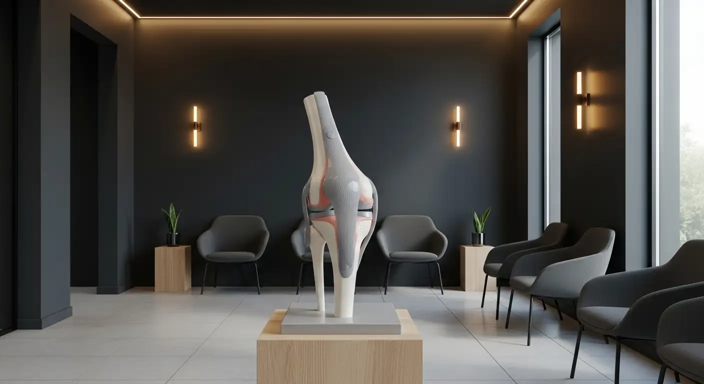
Understanding the Deep Infrapatellar Bursa
The deep infrapatellar bursa is a vital anatomical structure located in the knee region, playing a significant role in reducing friction and facilitating smooth movement of the knee joint. This article delves into its anatomical positioning, function, associated conditions, and the diagnostic approaches used to assess its health. For medical professionals, students, and individuals keen on understanding knee-related issues, this comprehensive overview offers valuable insights into the mechanisms and therapeutic interventions related to the deep infrapatellar bursa, enhancing our knowledge and clinical practice.
Anatomy and Function of the Deep Infrapatellar Bursa

Location and anatomical structure
The deep infrapatellar bursa is a significant anatomical structure located beneath the patellar tendon and just proximal to its insertion on the tibial tubercle. It lies directly posterior to the distal 38% of the patellar tendon, ensuring that it is well-positioned to cushion the areas that experience stress during knee movements. This bursal sac, part of the larger infrapatellar bursa system, is composed of two compartments, anterior and posterior, separated by a retropatellar fat pad. The average width of the deep infrapatellar bursa is about 30 mm, making it slightly wider than the patellar tendon, which assists in its identification during diagnosis and treatment procedures.
Function in reducing friction
The primary function of the deep infrapatellar bursa is to reduce friction between the patellar tendon and the tibia during knee flexion and extension. By producing synovial fluid, the bursa minimizes wear and tear on the tendon and surrounding tissues, thus facilitating smoother movement of the knee joint. This becomes particularly important during activities involving repetitive knee motion, like running and jumping, where friction levels can significantly increase.
Associated musculoskeletal movements
Movement patterns that commonly affect the deep infrapatellar bursa include activities that involve extensive knee flexion, such as deep squats and kneeling. These actions place added stress on the bursa, making it susceptible to conditions like deep infrapatellar bursitis, particularly in individuals engaged in repetitive jumping or those who frequently kneel, such as clergymen. It is crucial to understand the bursa's functional role to grasp the implications of its inflammation or injury, which can lead to substantial limitations in mobility and increased pain during everyday activities.
| Topic | Description | Importance in Clinical Settings |
|---|---|---|
| Location | Below the patellar tendon, proximal to the tibial tubercle. | Guides diagnosis and treatment options. |
| Function | Reduces friction during knee joint movement. | Essential for maintaining knee health and preventing injuries. |
| Associated Movements | Kneeling, running, jumping. | Understanding movement helps in prevention and rehabilitation strategies. |
Clinical Presentation and Diagnosis of Infrapatellar Bursitis

What are the symptoms of infrapatellar bursitis?
Symptoms of infrapatellar bursitis primarily include localized swelling just below the kneecap, which may feel soft and tender, often developing into a cyst from fluid accumulation due to trauma or infection. Patients typically report knee pain that radiates around the joint, worsening with activities such as climbing stairs or kneeling. Other symptoms that may accompany this condition include:
- Redness and Warmth: Skin over the inflamed bursa may become red and warm, particularly in septic cases.
- Stiffness or Rigidity: Patients might experience limited range of motion in the knee due to soft tissue damage surrounding the bursa.
- Systemic Symptoms: In severe instances, fever could arise if the bursitis is due to an infection, indicating the need for medical evaluation.
What are the imaging techniques used to assess deep infrapatellar bursa?
To accurately diagnose deep infrapatellar bursitis, imaging techniques play a critical role. Two primary modalities are:
| Imaging Technique | Description | Findings |
|---|---|---|
| Ultrasound | Utilizes sound waves to visualize soft tissue structures. | May reveal a distended bursa, cystic mass, and internal septations. |
| MRI | Employs magnetic fields to produce detailed images of the knee. | Displays bursal distension, synovial thickening, with T2-weighted images showing hyperintense signal and T1-weighted images appearing hypointense. |
These imaging techniques are essential not only for diagnosing deep infrapatellar bursitis but also for evaluating any associated conditions such as synovitis of the knee joint.
Differentiation from other knee conditions
It is crucial to differentiate infrapatellar bursitis from other knee pathologies such as patellar tendonitis, Osgood-Schlatter disease, and synovitis. While both infrapatellar bursitis and patellar tendonitis can cause anterior knee pain, infrapatellar bursitis specifically leads to swelling just below the kneecap, whereas tendonitis often presents more pain at the patellar tendon insertion. Additionally, conditions like Osgood-Schlatter disease are characterized by growth-related issues and may involve tenderness at the tibial tuberosity, rather than fluid accumulation in the bursa. Accurate diagnosis necessitates a thorough clinical evaluation and appropriate imaging to guide effective treatment.
Causes, Risk Factors, and Complications of Infrapatellar Bursitis

What causes deep infrapatellar bursitis?
Deep infrapatellar bursitis often arises from a combination of factors, including:
- Repetitive stress: Activities involving running, jumping, or prolonged kneeling can lead to excessive friction against the bursa.
- Acute trauma: Direct blows or falls onto the knee can trigger inflammation.
- Infections: Bacterial infections can cause septic bursitis, a form of inflamed bursa.
What conditions are associated with infrapatellar bursitis?
Deep infrapatellar bursitis can occur alongside other conditions, such as:
- Juvenile idiopathic arthritis
- Osgood-Schlatter disease
- Ankylosing spondylitis
- Patellar tendonitis These associations highlight the complexity of knee-related injuries and conditions.
What complications may arise from this condition?
Though often manageable, deep infrapatellar bursitis can lead to:
- Limited mobility: Pain during activities like kneeling or bending may significantly affect function.
- Chronic pain: If not addressed, inflammation can persist, leading to ongoing discomfort.
- Surgical considerations: Chronic cases may result in the need for surgical intervention if conservative treatments fail.
Treatment Approaches for Infrapatellar Bursitis

How is infrapatellar bursitis treated?
Treatment for infrapatellar bursitis mainly combines self-care measures and medical assistance. Initially, patients are advised to rest the affected knee and minimize activities that exacerbate pain. The acronym RICE—Rest, Ice, Compression, and Elevation—serves as a foundational approach. Applying ice helps reduce swelling, while gentle compression can provide support.
Medications commonly recommended include over-the-counter nonsteroidal anti-inflammatory drugs (NSAIDs) to alleviate pain and inflammation. If symptoms persist, physical therapy becomes essential. Therapists focus on strengthening the muscles surrounding the knee, which can help prevent future injuries.
In cases where the bursitis is caused by an infection, healthcare providers may prescribe antibiotics. Additionally, corticosteroid injections can be beneficial for reducing inflammation in the bursa directly.
Are there surgical interventions for infrapatellar bursitis?
Surgical options come into play only in more severe cases where conservative treatments fail to provide relief. Surgical removal of the inflamed bursa may be warranted, especially if it significantly interferes with daily activities or poses infection risks.
What is the role of physical therapy?
Physical therapy plays a pivotal role as part of the rehabilitative process after initial treatment. It typically focuses on restoring mobility and strength. Therapists employ exercises designed to improve knee function while also addressing any tightness or weakness, ultimately fostering better knee stability and reducing the chance of recurring bursitis.
Overall, effective management of infrapatellar bursitis involves a multidisciplinary approach, emphasizing individual care and tailored recovery plans.
Research Insights and Case Studies on Deep Infrapatellar Bursa

Noteworthy research findings
Research on the deep infrapatellar bursa has highlighted its role in various knee conditions, notably its involvement in bursitis cases. Studies, including dissected cadaveric knees, provide critical anatomical details, demonstrating the bursa's consistent positioning behind the patellar tendon and isolation from the knee joint. This anatomical knowledge is essential for diagnosing anterior knee pain accurately and understanding its clinical implications.
Case study analysis
One illustrative case involves a 40-year-old female presenting persistent knee pain and swelling localized to the infrapatellar bursa. Ultrasound imaging uncovered significant fluid accumulation, indicative of a ruptured bursa, while also identifying a Baker's cyst. Conservative treatment with anti-inflammatory drugs led to symptom resolution within six weeks, exemplifying effective management strategies for bursitis.
Clinical implications
Clinically, recognizing deep infrapatellar bursitis can prevent misdiagnosis, particularly in adolescents where symptoms may mimic Osgood-Schlatter disease. Recovery from infrapatellar bursitis typically takes around 2 to 6 weeks, influenced by the severity of the condition. Milder cases may resolve quickly; however, chronic issues necessitate more extensive interventions such as physical therapy and pain management strategies to ensure a full recovery.
Conclusion: Navigating the Deep Infrapatellar Bursa
Understanding the deep infrapatellar bursa is crucial for diagnosing and managing knee-related conditions effectively. With advances in imaging and therapeutic strategies, medical professionals can improve outcomes for patients experiencing disorders of this vital bursal structure. Continued research and case studies enrich our comprehension and capability to address the challenges posed by infrapatellar bursitis, empowering healthcare providers to optimize patient care and enhance recovery processes.
References
- Deep infrapatellar bursitis | Radiology Reference Article - Radiopaedia
- Infrapatellar Bursitis - Causes & Best Treatment Options in 2024
- Infrapatellar bursitis - Wikipedia
- [PDF] The Anatomy of the Deep Infrapatellar Bursa of the Knee
- Deep Infrapatellar Bursa | Complete Anatomy - Elsevier
- Deep infrapatellar bursa | Radiology Reference Article - Radiopaedia
- The anatomy of the deep infrapatellar bursa of the knee - PubMed
- Infrapatellar Bursitis | Twin Boro Physical Therapy - New Jersey
- Deep & Superficial Infrapatellar Bursitis: Symptoms & Treatment


