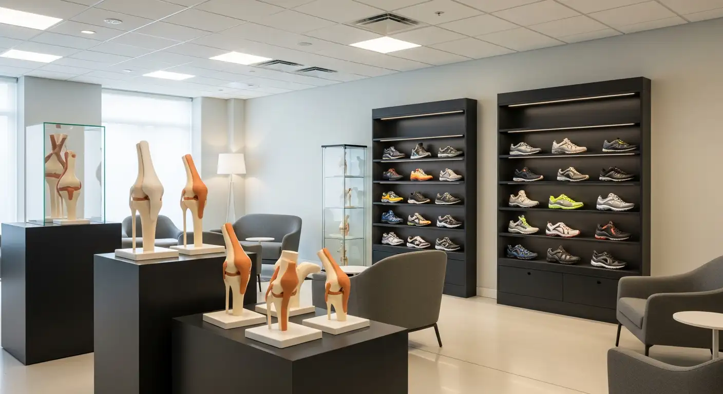
Understanding Knee Pain
Knee pain is a common issue that can affect individuals of all ages and activity levels. Understanding the causes, symptoms, and overall impact of knee pain is essential for addressing issues like fibula popping out of place at the knee.

Causes and Symptoms
There are various causes of knee pain that can stem from injuries, chronic conditions, or structural abnormalities. One specific issue that may arise is proximal tibiofibular ligament instability. This condition can manifest with symptoms such as pain, instability, and the sensation of the fibula popping out of place at the knee [1].
The following table outlines some common causes of knee pain and their associated symptoms:
CauseSymptomsLigament sprainsSwelling, pain, and instabilityMeniscus tearsLocking, popping, and swellingOsteoarthritisStiffness, pain during activity, and swellingProximal tibiofibular instabilityPain, instability, fibula popping sensationFracturesSudden onset of pain, inability to bear weight
Impact on Daily Life
Knee pain can significantly affect daily life. Individuals may struggle with common activities such as walking, climbing stairs, or participating in sports. Chronic knee pain can lead to reduced mobility, which may also affect mental health and overall quality of life.
In particular, conditions like proximal tibiofibular joint dislocation, although rare (accounting for only 1% of all knee injuries), can be easily overlooked during initial assessments [2]. Symptoms such as lateral knee pain, pressure over the fibular head, limited knee extension, and the sensation of popping may go unrecognized without proper clinical evaluation [3].
Seeking timely diagnosis and treatment is vital to prevent potential complications such as chronic pain or peroneal nerve palsy. Nonoperative treatment is often effective, but some individuals may require surgical intervention if they experience persistent instability [2].
Understanding these factors can help inform the approach to managing knee pain, particularly regarding specific conditions related to fibula instability.
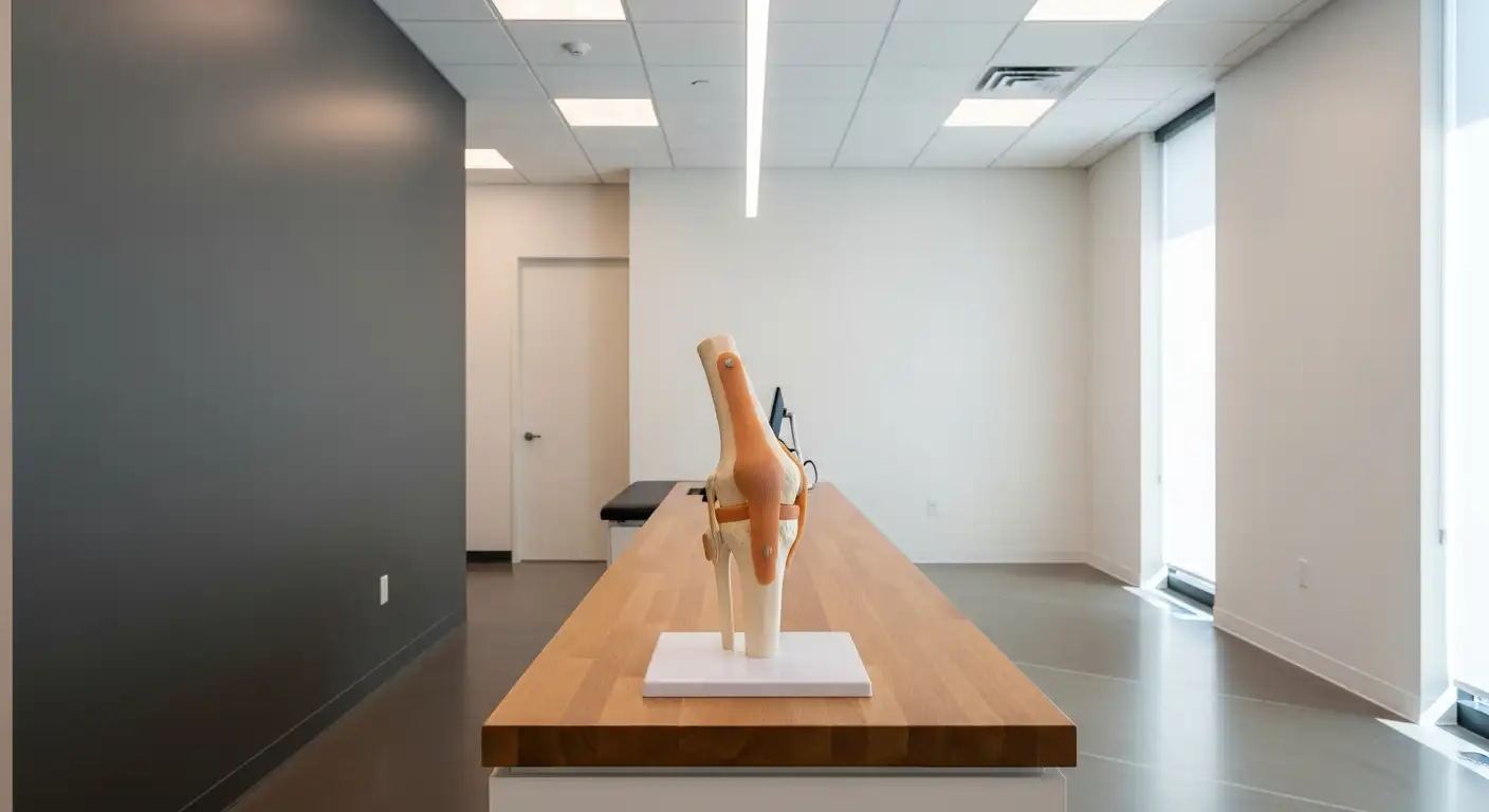
Fibula Dislocation at the Knee
Understanding fibula dislocation at the knee is essential for recognizing its symptoms and treatment options. One of the key issues associated with this condition is proximal tibiofibular ligament instability.
Proximal Tibiofibular Ligament Instability
Proximal tibiofibular ligament instability occurs when the ligaments stabilizing the proximal tibiofibular joint become compromised. This condition presents with symptoms such as pain, instability, and the sensation of the fibula popping out of place at the knee [1]. The instability can significantly impact mobility and day-to-day activities.
The condition is rare, accounting for only 1% of all knee injuries. Due to its low incidence, inadequate screening can cause it to be overlooked during initial assessments. Understanding the symptoms, such as recurrent knee pain and the characteristic popping sensation, is crucial for effective diagnosis and treatment.
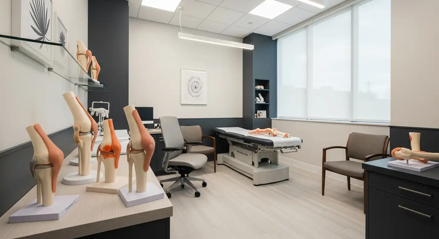
Diagnosis and Management
Diagnosing proximal tibiofibular ligament instability involves a detailed physical examination and careful review of imaging studies. A definitive diagnosis may be challenging, as this type of injury can be easily missed during routine evaluations [2].
Medical professionals may utilize the following diagnostic procedures:
Diagnostic MethodDescriptionPhysical ExaminationA thorough assessment to identify signs of instability and knee function.Imaging StudiesX-rays or MRI scans to visualize joint structure and rule out other injuries.
Once diagnosed, management strategies can be tailored. Nonoperative treatment options such as physical therapy may be effective for some individuals. However, studies indicate that approximately 50% of patients experiencing nonoperative treatment retain some level of instability, potentially leading to the need for surgical stabilization [2].
In cases where symptoms include chronic pain or complications like peroneal nerve palsy, surgical interventions, such as the use of a cortical button suspension device, may be warranted [2]. Early detection and prompt treatment are essential for improving outcomes and preventing long-term complications.
For those experiencing knee pain, it is important to seek professional evaluation to explore potential causes, including conditions like biceps femoris tendinopathy or other knee-related issues. Effective management can significantly enhance quality of life and mobility.
Fibula Fractures
Fibula fractures are breaks in the calf bone that commonly occur due to injuries like rolled ankles, awkward landings, falls, or direct blows to the leg. These types of fractures are prevalent in sports like football, basketball, and soccer due to the physical activities involved.
Types and Symptoms
Fibula fractures can be classified into two main types: open and closed. Understanding these types aids in identifying symptoms and determining the appropriate treatment.
Type of FractureDescriptionOpen FractureThe bone is exposed through the skin, or there is a deep wound exposing the bone. This type often requires more complex care due to the risk of infection.Closed FractureThe bone remains intact without breaking through the skin, but the bone is still fractured.
Symptoms of a fractured fibula generally include:
Treatment Options
Treatment approaches for fibula fractures depend on the fracture type and severity. Here are common treatment strategies:
Research suggests that after one to six weeks of nonoperative management, approximately 59% of patients with proximal tibiofibular joint dislocation improved without residual symptoms.
For related issues, such as biceps femoris tendinopathy that may arise alongside fibula injuries, and treatments such as the Don Joy knee brace for joint support, it's crucial to consult with a healthcare professional for personalized advice. Additionally, understanding your range of motion from studies on knee extension degrees can help guide recovery.
Proximal Tibiofibular Joint Dislocations
Rarity and Causes
Proximal tibiofibular joint dislocation is a very rare knee injury, accounting for only 1% of all knee injuries. This type of dislocation is most commonly caused by direct, high-energy trauma, such as from sports activities or traffic accidents. Isolated dislocations are often seen in sports that involve aggressive twisting of the knee, including soccer, parachuting, snowboarding, the long jump, horse riding, or scenarios involving direct trauma. There are four main injury patterns associated with this condition:
Dislocation TypePercentage of CasesMechanism of InjuryAnterolateral Dislocation85%Twisting motions, fallsPosteromedial Dislocation10%Plantar-flexed ankleSuperior Dislocation2%Direct trauma or extreme twisting
An injury to the proximal tibiofibular joint can cause the proximal fibula to sublux (partial dislocation) out of place over the lateral aspect of the knee joint, usually under stress during a fall or landing with the ankle plantar-flexed [4].
Diagnosis Challenges
The diagnosis of a proximal tibiofibular joint dislocation can be difficult to establish and is often missed during initial evaluation, even with plain radiographic examinations of the knee. Symptoms may include lateral knee pain, which worsens with pressure over the fibular head, limited knee extension, and crepitus or visual deformity. Diagnosis is substantially clinical, with findings such as locking or popping in the joint. Ankle movement may also exacerbate knee pain, and transient peroneal nerve palsy may occur with posterior or superior dislocations [3].
Plain radiography has shown inconsistencies in aiding the diagnosis. Therefore, computed tomography (CT) is recommended as the investigation of choice when this injury is suspected, as it provides a clearer assessment of the congruity of the proximal tibiofibular joint. Careful evaluation, particularly on the lateral view of the knee, is essential for diagnosis [3].
Early detection and prompt treatment are essential to prevent complications such as chronic pain and peroneal nerve palsy. Issues like biceps femoris tendinopathy can arise if not treated properly, underscoring the importance of being vigilant in the presence of knee pain.
Surgical Treatments
When conservative methods fail to restore stability and function in cases of fibula popping out of place at the knee, surgical intervention may become necessary. Two key components of surgical treatment are anatomical reconstruction and the rehabilitation process.
Anatomical Reconstruction
Surgical procedures for addressing proximal tibiofibular joint instability often involve an anatomical reconstruction of the torn ligaments, particularly the posterior proximal tibiofibular joint ligaments. This reconstruction is typically executed using grafts, which can be either hamstring allografts or autografts. The grafts are positioned in an anatomically correct manner to ensure optimal stabilization of the joint.
In acute cases, the initial treatment may include immobilization in a brace for three weeks. However, if the injury is chronic, surgical reconstruction may be required to restore stability effectively. The surgical technique aims to realign the fibula and restore its proper function within the knee joint.
Type of ReconstructionDescriptionAllograftGraft from a donorAutograftGraft taken from the patient's own body
Rehabilitation Process
Post-surgical rehabilitation plays a crucial role in recovery after fibula stabilization surgery. Initially, patients are kept non-weight bearing for approximately six weeks. During this time, the focus is on allowing the reconstructed ligaments to heal properly. After the initial healing phase, patients typically engage in a progressive rehabilitation program that may include:
Physical therapy becomes a vital part of recovery, focusing on restoring range of motion, strength, and stability in the knee. Effective rehabilitation can significantly improve the chance for full recovery and return to normal activities.
Proper execution of these surgical and rehabilitation protocols can greatly enhance outcomes for individuals experiencing instability due to fibula popping out of place at the knee. For further information on knee pain management, including related conditions like biceps femoris tendinopathy, various braces such as the Don Joy knee brace, or related knee mechanics like knee extension degrees, be sure to explore the linked resources.
Importance of Early Detection
Detecting issues related to fibula dislocation at the knee early can significantly influence treatment outcomes and the quality of life for individuals affected. Understanding prevention strategies and the long-term implications of this condition is essential.
Prevention Strategies
Taking proactive measures can help mitigate the risk of proximal tibiofibular ligament instability and related injuries. Here are some key strategies:
StrategyDescriptionStrengthening ExercisesFocusing on exercises that strengthen the muscles surrounding the knee can provide better support and stability.Balance TrainingEngaging in balance training can improve coordination and prevent falls that may lead to injuries.Proper FootwearWearing appropriate shoes can help absorb impact and provide adequate support during activities.Warm-up and StretchingIncorporating warm-up routines and stretching before physical activities can reduce the risk of injury.
Individuals should consult with a healthcare professional for personalized recommendations on exercises and preventive measures.
Long-term Implications
Failing to recognize and adequately treat fibula dislocation at the knee can lead to several long-term complications:
ComplicationDescriptionChronic PainProlonged instability can lead to ongoing discomfort and affect daily activities.Peroneal Nerve PalsyDamage to the peroneal nerve may occur, resulting in weakness or numbness in the foot.Joint InstabilityContinuous instability can result in repeated dislocations or subluxations, requiring surgical intervention.Compromised MobilityChronic pain and instability can limit physical activity, leading to a sedentary lifestyle.
Early identification of symptoms such as pain, instability, or the sensation of the fibula popping out of place at the knee is critical. Prompt treatment can help prevent these long-term issues and improve overall health outcomes.
For more information about knee injuries, including biceps femoris tendinopathy and the use of supports like a Don Joy knee brace, interested individuals are encouraged to explore further resources. Additionally, understanding knee extension degrees can be beneficial for individuals working on rehabilitation or preventive strategies.
References
[2]:
[3]:
[4]: