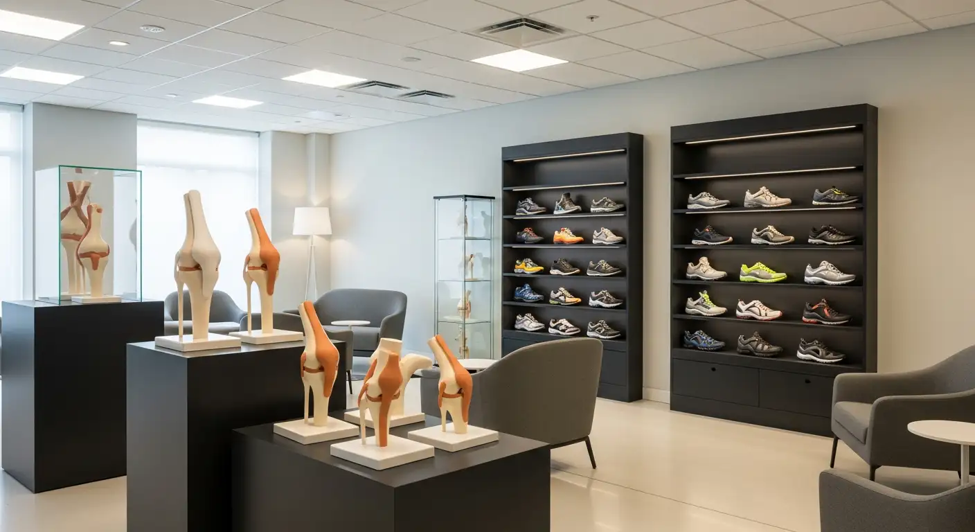
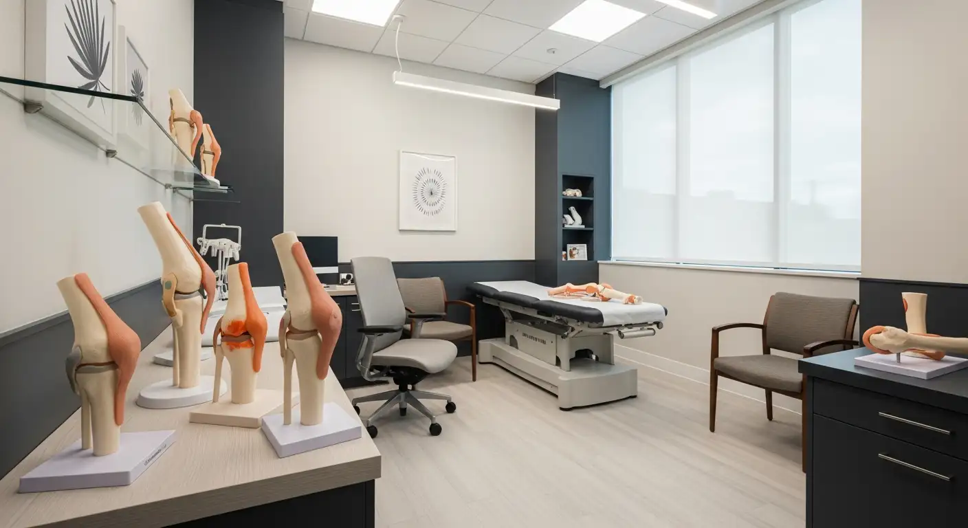
Understanding the Patella
The patella, often referred to as the knee cap, plays a crucial role in the function and movement of the knee. Let's delve into the anatomy and function of the patella to better understand its importance.
Anatomy of the Patella
The patella is the largest sesamoid bone in the body, typically shaped like an upside-down triangle. In terms of dimensions, it typically measures 4 - 4.5 centimeters in length, 5 -5.5 centimeters in width, and 2 - 2.5 centimeters in thickness [1].
Interestingly, the patella bone only forms between the ages of three and six years old. Some individuals may even have what's known as a bipartite patella, where the bone forms as two separate entities.

In humans, as well as in other studied organisms like mice, rabbits, and chicks, the patella is not yet ossified at birth. The ossification process typically begins at about 5-6 years of age and completes during adolescence. This bone develops within the quadriceps femoris tendon, classifying it as a true sesamoid bone.
Function of the Patella
The primary function of the patella is to aid in knee extension and movement. By increasing the angle of the quadriceps tendon as it pulls on the lower leg, the patella enhances the mechanical advantage of this muscle group and thus facilitates knee extension.
At full knee extension, the patella provides 31% of total knee extension torque. However, between 90 and 120 degrees of flexion, it only contributes about 13%.
In addition to aiding in movement, the patella also provides protection for the knee joint. It acts as a bony shield for the anterior trochlea, preventing excessive friction between the quadriceps tendon and the femoral condyles [1].
Understanding the anatomy and function of the patella is crucial for comprehending various knee-related conditions and treatments, including those associated with high knee caps. The following sections will delve into these in more detail.
Common Patella Conditions
The patella, or kneecap, can be prone to several conditions that can affect mobility and cause discomfort or pain. Understanding these conditions is the first step in seeking appropriate treatment.
Patella Alta and Patella Baja
The patella's position in relation to the knee joint can significantly influence its function. When the patella is high-riding, or superiorly aligned, it is referred to as Patella Alta. An attenuated Patella Alta is an unusually small kneecap that develops out of and above the joint. On the other hand, a low-riding patella is known as Patella Baja. A long-standing Patella Baja may result in extensor dysfunction.
These conditions can affect the knee's function, as the main role of the patella is to aid knee extension and movement, while also shielding the knee joint [2].
Patellar Tracking Disorder
Patellar Tracking Disorder is a condition where the kneecap does not stay in place as the leg bends and straightens. This abnormal movement can lead to discomfort and knee instability.
The thigh muscles, or quadriceps, play a crucial role in keeping the kneecap stable. Weak quadriceps can increase the risk of Patellar Tracking Disorder. The stability of the patella is also aided by ligaments and tendons. If these are too tight or too loose, the likelihood of Patellar Tracking Disorder rises.
Patellar Tendonitis
Patellar Tendonitis, also known as Jumper's Knee, is a common condition related to the patella. It often occurs in individuals who are very active or participate in sports involving jumping.
In this condition, the tendon connecting the kneecap to the shinbone becomes inflamed due to overuse or injury. This can cause pain and tenderness around the patella, making it difficult to perform activities that put pressure on the knee, such as running and jumping.
These are just some of the common conditions that can affect the patella. If you experience knee pain or notice changes in the function or appearance of your kneecap, it is important to seek medical advice to ensure appropriate diagnosis and treatment.
Treatment and Rehabilitation
When it comes to issues related to high knee caps or patella problems, the path to recovery often lies in effective treatment and diligent rehabilitation. This journey could involve non-surgical treatment options, specific exercises, and tailored rehabilitation programs.
Non-Surgical Treatment Options
The goals of non-surgical treatment for conditions like patellar tracking problems are to reduce symptoms, increase quadriceps strength and endurance, and return to normal function. Often, straightforward exercises for patellar tracking disorder can be performed at home, requiring only about 20 minutes a day. Such a routine, while simple, can lead to significant improvements over time.
Exercises for Patellar Tracking Disorder
Exercise plays a crucial role in the non-surgical treatment of conditions like patellar tracking disorder. Basic exercises such as quad sets and mini squats may be the initial recommendations from your doctor or physiotherapist. These exercises aim to strengthen the quadriceps, the muscle grouping at the front of your thigh, which plays a crucial role in knee stability and movement.
Tailored Rehabilitation Programs
Each individual's knee condition is unique, and thus, a one-size-fits-all approach to rehabilitation is rarely effective. Correct diagnosis is critical in designing an effective exercise or rehabilitation program. The treatment received and the exercise program used to rehabilitate the knee should be developed specifically for the individual's condition. By tailoring the program to the individual's needs, the chances of a successful recovery are significantly increased.
By combining non-surgical treatment options, specific exercises, and personalized rehabilitation programs, it's possible to effectively manage conditions related to high knee caps. Remember to consult with a healthcare professional before beginning any new exercises or treatment plans to ensure safety and effectiveness.
Impact of Patella on Knee Health
The position and health of the patella, also known as the kneecap, significantly impact overall knee health. This section will focus on patellar dislocations, recurrent patellar dislocations, and the anatomical factors involved in these conditions.
Patellar Dislocations
Patellar dislocations occur when the kneecap moves out of its normal position, often caused by a sudden twist or direct impact. This condition is characterized by intense knee pain, inability to straighten the knee, and visible dislocation of the kneecap. Immediate medical attention is recommended to prevent further complications.
Recurrent Patellar Dislocations
Recurrent patellar dislocations refer to repeated instances of patellar dislocation. This condition is related to the anatomical alignment and imbalance of bone and soft tissue, with adolescents with immature bones being particularly susceptible. According to NCBI, the incidence of ipsilateral recurrence after the first attack is 36%, and contralateral dislocation is 5%.
Anatomical Factors in Patellar Dislocations
Several anatomical factors contribute to patellar dislocations. Abnormal anatomical structure of the knee joint, such as significant pulley dysplasia, is identified as the main cause of patellar dislocation. A study cited by NCBI reported that the trochlear angle in patients with recurrent patellar dislocation was measured to be 16.0 ± 3.9°, the trochlear sulcus angle was 165.8 ± 8.7°, and the trochlear sulcus depth was 1.54 ± 1.25 mm.
Anatomical measurements commonly evaluated in patients with recurrent patellar dislocation include trochlear dysplasia classification, trochlear angle, trochlear sulcus angle, trochlear sulcus depth, Insall-Salvati index, Caton-Deschamps index, tibial tubercle–trochlear groove distance, patellar tilt, and Q angle.
Understanding these anatomical factors can aid in the diagnosis and treatment of patellar dislocations, potentially reducing the likelihood of recurrent dislocations and improving knee health.
Biomechanics of the Knee
Understanding the biomechanics of the knee is fundamental to grasping the impact of high knee caps on knee health. This section will explore the vulnerability of the knee joint, common knee musculoskeletal disorders, and the role of knee osteoarthritis in these issues.
Knee Joint Vulnerability
The knee joint plays an essential role in our daily lives, supporting our body weight, assisting with the swing of our lower limbs, and absorbing the shock of our movements. It's capable of withstanding tremendous forces during normal movements, making it a crucial area of study for human locomotor function assistance or rehabilitation. However, these frequent and intense pressures also make the knee joint susceptible to various impairments.
Knee Musculoskeletal Disorders
Knee musculoskeletal disorders, such as knee osteoarthritis (KOA), significantly affect knee biomechanics. Interestingly, KOA is more commonly observed in the medial (inner) compartment of the knee than the lateral (outer) compartment, with the medial compartment bearing about 70% of the total force. This disproportionate load distribution can lead to the progression of musculoskeletal disorders, impacting the overall health and function of the knee.
Knee Osteoarthritis
Knee osteoarthritis (KOA) is a prevalent condition affecting the biomechanics of the knee, leading to pain and mobility issues. In KOA, the medial compartment of the knee bears about 70% of the total force, leading to a higher prevalence of medial KOA compared to lateral KOA. Genu varum alignment, a condition where the knees bow outwards, can increase the load on the medial compartment, thereby impacting the progression of the disease.
By understanding the biomechanics of the knee and factors that contribute to knee disorders, healthcare professionals can develop effective treatment plans to manage these conditions. Such understanding also underscores the need for preventative measures, such as proper exercise and body mechanics, to maintain knee health, especially in individuals with high knee caps.
Genetic Factors in Patellar Dislocations
Understanding the genetic factors in patellar dislocations can shed light on why some individuals may be more susceptible to these types of injuries. Research in this area is ongoing, but some discoveries have already been made.
Genes Associated with Patellar Dislocations
Genetic research into patellar dislocations has identified two potentially significant genes: HOXB9 and SLC26A2. Whole exome sequencing revealed mutations in these genes that were only present in samples from patients with recurrent patellar dislocation. This suggests that these genes could be pathogenic or related to dislocation of the patella.
Anatomical Measurements in Recurrent Dislocations
In addition to genetic factors, anatomical measurements can also play a role in recurrent patellar dislocations. Measurements taken from patients with recurrent dislocations showed a trochlear angle of 16.0 ± 3.9°, a trochlear sulcus angle of 165.8 ± 8.7°, and a trochlear sulcus depth of 1.54 ± 1.25 mm. These measurements indicate significant pulley dysplasia, an abnormality that may contribute to recurrent dislocations. NCBI
Abnormal Anatomical Structures
Abnormal anatomical structures of the knee joint have also been identified as a primary cause of patellar dislocation. These abnormalities include variations in the trochlear dysplasia classification, trochlear angle, trochlear sulcus angle, trochlear sulcus depth, Insall-Salvati index, Caton-Deschamps index, tibial tubercle–trochlear groove distance, patellar tilt, and Q angle.
Understanding the genetic and anatomical factors that contribute to patellar dislocations can help medical professionals develop more effective prevention and treatment strategies. As research continues to evolve in this area, even more insights into the causes and management of patellar dislocations are likely to emerge.
References
[1]: https://www.ncbi.nlm.nih.gov/pmc/articles/PMC5095937/
[2]: https://www.verywellhealth.com/patella-anatomy-function-and-treatment-4768658
[3]: https://www.ncbi.nlm.nih.gov/pmc/articles/PMC6461774/
[4]: https://en.wikipedia.org/wiki/Patella
[5]: https://www.healthlinkbc.ca/health-topics/patellar-tracking-disorder-exercises