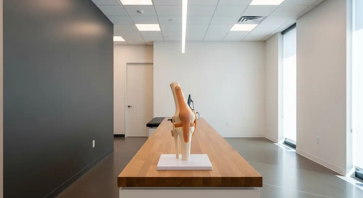
Understanding Knee Anatomy
The knee joint comprises several key structures that play crucial roles in its function and stability. Among these, bursae and ligaments are essential components.
Bursae and Their Function
Bursae are small, fluid-filled sacs located in various joints of the body. They serve to reduce friction between moving parts, thereby lessening wear and tear on the joint. In the knee, bursae cushion pressure points between bones and tendons, muscles, and skin [1].
Bursae around the knee can be categorized into two groups: those located near the kneecap (patella) and those positioned elsewhere. The following table lists the bursae associated with the knee:
Bursa TypeLocationPrepatellar BursaIn front of the kneecapSuperficial Infrapatellar BursaBelow the kneecap's surfaceDeep Infrapatellar BursaBehind the kneecapSuprapatellar BursaAbove the kneecapPes Anserine BursaMedial aspect of the tibiaIliotibial BursaLateral aspect of the kneeGastrocnemius-Semimembranosus BursaNear the calf muscles
Understanding these bursae is critical, as conditions like knee bursitis can arise from their inflammation or swelling, leading to pain and irritation [2].
Ligaments in the Knee
Ligaments are tough, flexible bands of connective tissue that connect bones to other bones. In the context of the knee, the lateral collateral ligament (LCL) is particularly significant. It connects the femur to the fibula, providing stability against lateral movement of the knee. The LCL plays a role in controlling sideways motion and braces the knee against unusual movement [3].
The function of the LCL can be summarized as follows:
FunctionDescriptionStabilization against lateral movementControls side-to-side motion to prevent injuryVarus stress resistanceBraces the knee during varus (bow-legged) stressPosterolateral rotation controlHelps in stabilizing rotation of the tibia relative to the femur
The LCL acts as a secondary stabilizer when the cruciate ligaments are compromised, preventing excessive anterior and posterior movement of the tibia [4].
Understanding the anatomy of the knee, including its bursae and ligaments, is fundamental for recognizing potential issues and addressing knee pain effectively. For further insights on knee injuries and management, consider exploring knee bends and lateral release knee options.
The Role of Menisci
Menisci are essential components of the knee joint, playing a vital role in its function and stability. The knee contains two menisci: the medial meniscus and the lateral meniscus. Each provides unique benefits that contribute to overall knee health.
Function of Medial Meniscus
The medial meniscus is positioned on the inner side of the knee and has several key functions:
In summary, the medial meniscus is crucial for maintaining knee functionality and stability, especially during high-impact movements.
Function of Lateral Meniscus
The lateral meniscus serves a similar purpose but is located on the outer side of the knee. Its functions include:
The lateral meniscus is made of tough, smooth, and rubbery fibrocartilage, forming a C-shape. It is physically attached to the tibia (shin bone) and comes into contact with the femur (thigh bone). By improving joint stability and distributing weight more evenly, both menisci play a critical role in maintaining knee health.
Understanding lateral knee anatomy is essential for recognizing knee pain's causes and developing effective treatment strategies. For further information on common knee injuries, consider exploring our resources on knee brace for ACL tear and patellar maltracking.
Exploring the Lateral Meniscus
The lateral meniscus is a critical component of knee anatomy, providing stability and cushioning to the joint. This section discusses the structure and composition of the lateral meniscus as well as its nourishment and blood supply.
Structure and Composition
The lateral meniscus is shaped like a C and consists of tough, smooth, rubbery fibrocartilage. It is attached to the top of the shin bone (tibia) and makes contact with the thigh bone (femur). This anatomical structure enables it to act as a shock absorber during weight-bearing activities, distributing body weight more evenly and enhancing joint stability.
Key features of the lateral meniscus include:
FeatureDescriptionShapeC-shapedMaterialFibrocartilageAttachmentsAttached to tibia; contacts femurFunctionShock absorption and joint stability
The lateral meniscus also has a wedged profile that helps in maintaining the stability of the knee joint by preventing the rounded femur surface from sliding off the flat tibial surface.
Nourishment and Blood Supply
The blood supply to the meniscus is crucial for its health and functionality. The lateral meniscus has an area known as the vascular zone, which contains small blood vessels that nourish the tissue. Conversely, it also has an avascular zone, which lacks direct blood supply. The red/white zone lies between these two areas and has minimal blood supply [5].
In summary, the blood supply within the lateral meniscus is as follows:
ZoneBlood SupplyVascular ZoneRich blood supplyRed/White ZoneMinimal blood supplyAvascular ZoneNo blood supply
Understanding the lateral knee anatomy, particularly the structure and nourishment of the lateral meniscus, is essential for recognizing its role in knee stability and for addressing potential injuries.
Lateral Collateral Ligament (LCL)
The lateral collateral ligament (LCL) plays an essential role in maintaining the stability of the knee joint.
Function of LCL
The LCL connects the femur to the fibula, controlling the sideways motion of the knee and bracing it against unusual movements [3]. It acts as the primary varus stabilizer of the knee and is crucial for stabilizing the joint during various activities. The LCL is particularly important in providing resistance to varus stress, which occurs when the knee is pushed inward.
Key functions of the LCL include:
In a fully extended knee, the LCL is actively engaged, whereas it becomes less taut when the knee is flexed beyond 30° [4].
FunctionDescriptionVarus StabilizationResists inward knee movement and maintains alignment.Control of RotationPrevents excessive rotation and instability during movement.
Injuries and Symptoms
Injuries to the LCL are most commonly the result of high energy trauma, such as a direct blow to the anteromedial aspect of the knee while it is extended, leading to hyperextension and excessive varus force. Common symptoms of LCL injury include:
It's important to note that LCL injuries are often accompanied by other knee ligament injuries, particularly those to the anterior cruciate ligament (ACL), posterior cruciate ligament (PCL), and the posterior-lateral corner (PLC) of the knee [6].
For those experiencing tightness in their knee, it may help to explore stretches or exercises targeting the related muscle groups. Additional techniques such as knee bends and gastroc stretches can be beneficial in maintaining flexibility and stability in the knee joint. If knee pain persists or worsens, consultation with a healthcare professional is advisable.
Iliotibial Band (ITB)
ITB Anatomy and Function
The iliotibial band (ITB) is a longitudinal fibrous sheath that runs along the lateral thigh and is crucial for lower extremity motion. It spans the lateral aspect of the lower body before inserting on Gerdy's tubercle on the proximal/lateral tibia [7]. The ITB plays a vital role in stabilizing the knee during activities such as running and walking.
This structure is composed primarily of collagen, allowing for both strength and flexibility. It helps to manage lateral stability and aids in the proper tracking of the knee joint, especially during dynamic movements.
FeatureDescriptionStructureLongitudinal fibrous sheathLocationLateral thigh to proximal/lateral tibiaCompositionCollagen fibersFunctionStabilizes knee, aids in lower limb motion
Iliotibial Band Syndrome (ITBS)
Iliotibial band syndrome (ITBS) is one of the leading causes of lateral knee pain among runners. It accounts for 5–14% of all running-related injuries, with a prevalence rate of 50-81% in male runners [8]. ITBS occurs when the ITB becomes tight or inflamed, often leading to pain on the outer side of the knee.
Symptoms of ITBS often include:
Management of ITBS typically involves rest, physical therapy, and specific exercises aimed at stretching and strengthening the hip and knee muscles. For more information about exercises that may help alleviate symptoms, see our articles on gastroc stretch, glute med stretch, and exercises for upper glutes.
Diagnosis and Treatment
Accurate diagnosis and effective treatment are essential for managing issues related to knee pain, particularly concerning lateral knee anatomy. Understanding the necessary diagnostic procedures and potential management strategies can facilitate appropriate care.
Diagnostic Procedures
Diagnosing knee injuries typically begins with a comprehensive physical examination. The physician will compare the injured knee with the uninjured one and assess the knee's range of motion and overall function. Various diagnostic imaging techniques may be utilized to confirm the injury and assess the extent of damage:
Diagnostic MethodDescriptionX-raysUseful for visualizing bone structures and identifying fractures.MRIProvides detailed images of soft tissues, including ligaments, tendons, and cartilage, essential for diagnosing ACL tears and meniscus injuries.UltrasoundCan be used to assess soft tissue injuries and joint conditions.
For conditions like ACL injuries and meniscus tears, confirmation through MRI is important for visualization and determining the best treatment pathway.
Management and Interventions
Treatment for knee injuries varies based on the specific diagnosis and severity. Common management strategies include:
Treatment OptionDescriptionPhysical TherapyA comprehensive approach that includes exercises to strengthen muscles around the knee, improve flexibility, and restore function.Knee BracingOffers support and stability during the healing process, particularly for ACL injuries. Options include custom knee braces and off-the-shelf varieties [9].Surgical InterventionsIn severe cases, surgical options such as ACL reconstruction or meniscus repair might be necessary. Techniques such as arthroscopy, the BEAR procedure, or lateral extra-articular tenodesis could be recommended (Plancher Orthopaedics).
In addition to these treatments, specific exercises can help improve overall knee health. Stretching routines like the gastroc stretch and glute med stretch can enhance flexibility, while targeted strength training, such as the vastus lateralis workout, can bolster knee stability.
Monitoring symptoms such as tightness or pain is crucial. If the individual experiences discomfort while performing activities like knee bends (knee bends) or going up and down stairs, strategies for relief should be pursued. Understanding these diagnostic and treatment options empowers individuals to seek appropriate care for their knee concerns, fostering better health outcomes.
References
[2]:
[3]:
[4]:
[5]:
[6]:
[7]:
[8]:
[9]: