Understanding Knee Anatomy

Structure of the Knee Joint
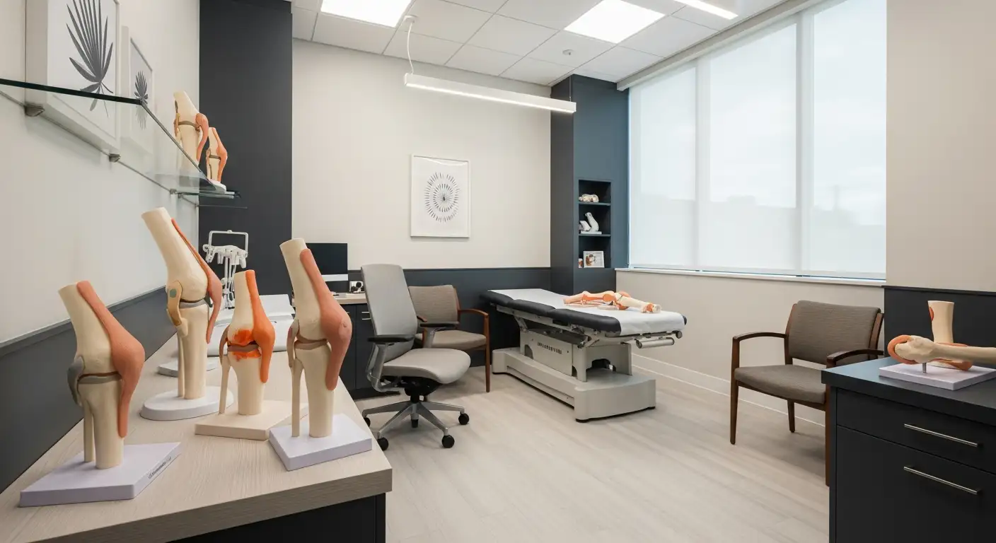
The knee joint, the largest joint in the body, functions as a synovial joint, allowing for a wide range of movement. It is where the thigh bone (femur) meets the shin bone (tibia) in the middle of the leg [1]. This hinge joint operates similarly to a door hinge, enabling flexion and extension. Key components of the knee joint include:
- Femur: The upper leg bone or thigh bone.
- Tibia: The larger bone in the lower leg.
- Patella: The kneecap, which protects the joint.
- Fibula: The smaller bone in the lower leg.
These bones are connected by a complex set of ligaments, tendons, and muscles, providing stability and facilitating movement. The knee joint is also cushioned by cartilage, including the medial and lateral menisci, which act as shock absorbers.
Function of the Knee Joint
The knee joint's primary function is to support the body's weight during activities such as standing, walking, running, and jumping. It allows for flexion (bending) and extension (straightening) of the leg, as well as slight internal and external rotation.
The medial meniscus, located on the inner side of the knee joint, and the lateral meniscus, positioned on the outer side, play crucial roles in maintaining knee stability and facilitating smooth movement [2]. They distribute the load across the knee, absorb shock, and reduce friction during movement.
Understanding the structure and function of the knee joint is essential for recognizing the importance of maintaining its health. For individuals seeking non-surgical treatments for knee osteoarthritis, options such as knee compression sleeves for swelling and knee injection sites may provide relief. Additionally, conditions like the tibial tuberosity bump in adults and issues with the lateral retinaculum can impact knee function and require proper evaluation and management.
Meniscus Tears
Meniscus tears are common injuries in the knee joint, particularly affecting the medial joint line. Understanding the types, symptoms, and diagnosis of meniscus tears is essential for individuals seeking non-surgical treatments for knee osteoarthritis.
Types of Meniscus Tears
Meniscus tears can vary widely in size and severity. They can be classified into several types based on their shape and location. According to Cedars-Sinai, common types include:
- Radial Tears: These tears occur perpendicular to the circumference of the meniscus.
- Horizontal Tears: These tears run parallel to the tibial plateau and are often associated with meniscal cysts.
- Bucket Handle Tears: These are vertical longitudinal tears that may cause the meniscus to displace into the joint.
- Flap Tears: These are oblique tears that create a flap of meniscal tissue.
- Complex Tears: These involve a combination of tear patterns.

Symptoms and Diagnosis
The symptoms of a meniscus tear can range from mild to severe, affecting the functionality of the knee. Common symptoms include:
- Severe pain and swelling that may develop up to 24 hours after the injury
- Difficulty walking
- Additional pain when flexing or twisting the knee
To diagnose a meniscus tear, healthcare providers often rely on clinical evaluations and imaging techniques.
Clinical Evaluation
A thorough clinical evaluation includes assessing the patient's history and conducting physical examinations. One commonly used test is the Joint Line Tenderness (JLT) test, which helps identify tenderness along the medial joint line, indicating a possible meniscus tear. The sensitivity and specificity of the JLT test can provide useful information for diagnosis.
Imaging Techniques
Magnetic Resonance Imaging (MRI) is a crucial tool for diagnosing meniscus tears. According to Cedars-Sinai, MRI findings classify meniscus tears into three grades:
Meniscal tears have a high prevalence, with a study in Switzerland reporting a point prevalence of 57% in symptomatic individuals and 36% in asymptomatic individuals. In the U.S., the prevalence is 32% in symptomatic individuals and 23% in asymptomatic individuals [3]. Each year, two out of a thousand people in the Netherlands are diagnosed with new meniscal tears.
For more information on managing knee pain and related conditions, explore our articles on lateral retinaculum, knee compression sleeve for swelling, and knee injection sites.
Medial Meniscus
The medial meniscus plays a critical role in the knee joint, providing stability, shock absorption, and aiding in the prevention of knee-related conditions such as osteoarthritis. Understanding its function and anatomy is essential for those seeking non-surgical treatments for knee osteoarthritis.
Role of the Medial Meniscus
The medial meniscus is a crescent-shaped, cartilaginous band located between the medial tibial and medial femoral condyle. It covers approximately 60% of the contact area of the medial compartment of the knee joint. The medial meniscus serves several key functions:
- Load Distribution: It helps to evenly distribute the load across the knee joint, reducing stress on the joint surfaces.
- Stabilization: It provides stabilization to the knee, particularly during rotational movements and changes in direction.
- Shock Absorption: It acts as a cushion to absorb shock and reduce the impact on the knee joint during activities such as walking, running, and jumping.
- Nourishment: It aids in the nourishment of the knee joint by facilitating the movement of synovial fluid.
- Proprioception: It contributes to proprioception, allowing the knee to sense its position and movement.
- Joint Gliding: It allows for smooth gliding of the joint surfaces, enhancing overall knee function.
- Prevention of Hyperextension: It prevents hyperextension of the knee, protecting it from injury.
- Osteoarthritis Prevention: It plays a role in preventing the development of knee osteoarthritis by distributing mechanical loads and providing cushioning.
Due to its firm attachment to the deep surface of the medial collateral ligament (MCL) medially, the anterior cruciate ligament (ACL) anteriorly, and the posterior cruciate ligament (PCL) posteriorly, the medial meniscus is less mobile than the lateral meniscus [5]. This limited mobility makes it susceptible to injury, particularly in activities involving twisting or sudden directional changes.

Vascular Supply and Nerve Innervation
The vascular supply of the medial meniscus is primarily derived from the medial, lateral, and middle genicular arteries, which are branches of the popliteal artery. Only 10% to 30% of the meniscus receives direct blood supply; the remaining portion is nourished through diffusion from synovial fluid or mechanical motion [4].
The medial meniscus is innervated by the articular branch of the posterior tibial nerve and the terminal branches of the obturator and femoral nerves. Various mechanoreceptors, including Type I (Ruffini), Type II (Pacinian), and Type III (Golgi tendon organ) mechanoreceptors, are present in the medial meniscus. These mechanoreceptors play a vital role in supporting movement and positional adaptation.
Understanding the intricate details of the medial meniscus, including its role, vascular supply, and nerve innervation, is crucial for comprehending the biomechanics and function of the knee joint. For more information on related structures, consider exploring our articles on the lateral retinaculum and knee injection sites.
Medial Supporting Structures
The medial joint line of the knee is supported by a complex network of structures that play a critical role in maintaining stability and function. Understanding these medial supporting structures is essential for those seeking non-surgical treatments for knee osteoarthritis.
Layers of Medial Supporting Structures
The medial supporting structures of the knee are divided into three distinct layers: superficial, intermediate, and deep.
- Superficial Layer:
- Deep crural fascia
- Intermediate Layer:
- Tibial collateral ligament (TCL)
- Deep Layer:
- Deep medial capsular ligament
- Joint capsule
The superficial layer consists of the deep crural fascia, providing a basic level of support. The intermediate layer primarily comprises the tibial collateral ligament (TCL), which is a band-like ligament with one femoral and two tibial attachments. The deep layer is formed by the deep medial capsular ligament and the joint capsule. The deep medial capsular ligament has two components: the meniscofemoral ligament and the meniscotibial ligament.
Function and Importance
The medial supporting structures are vital for the stability and proper function of the knee. Among these, the tibial collateral ligament (TCL), deep medial capsular ligament, and posterior oblique ligament (POL) are the most important static stabilizers.
- Tibial Collateral Ligament (TCL): This ligament provides significant medial stability and has an average tensile strength of 557 Newtons, highlighting its resilience.
- Deep Medial Capsular Ligament: This ligament, with an average tensile strength of 101 Newtons, acts as a thickening of the medial joint capsule.
- Posterior Oblique Ligament (POL): The POL contributes to the medial stability with an average tensile strength of 256 Newtons [6].
These structures work together to ensure the knee remains stable during various activities. Damage or weakening of any of these structures can lead to instability and pain, often necessitating medical intervention. For more information on knee anatomy, explore our articles on the lateral retinaculum and knee injection sites. Understanding the medial supporting structures is crucial for effective diagnosis and treatment of knee conditions, providing a foundation for non-surgical management options.
Joint Line Tenderness
Joint Line Tenderness (JLT) is a crucial diagnostic tool used to evaluate knee injuries, particularly meniscal tears. This section will explore the JLT test and its sensitivity and specificity.
Joint Line Tenderness Test
The Joint Line Tenderness (JLT) test is commonly used to screen for sensitivity related to meniscal injuries. It can be employed if pain is localized to either the medial or lateral aspect of the knee joint, correlating with degenerative pathology of the articular joint cartilage or compromised integrity of the medial or lateral meniscus [7].
How to Perform the JLT Test:
- The patient lies supine with the knee flexed at approximately 90 degrees.
- The examiner palpates along the medial and lateral joint lines of the knee.
- The test is considered positive if the patient experiences pain or tenderness upon palpation.
The JLT test is simple and non-invasive, making it a popular choice for initial clinical evaluations. For more information on knee anatomy and other diagnostic tests, visit our section on knee injection sites.
Sensitivity and Specificity
The sensitivity and specificity of the JLT test are generally high but vary between the medial and lateral meniscus. Studies have shown that the sensitivity for the medial meniscus is 86% with a specificity of 67%, while for the lateral meniscus, the sensitivity is 92% with a specificity of 97%.
While the JLT test has a high sensitivity, its relatively lower specificity indicates that patients with JLT may not exclusively have meniscal tears, especially medial meniscus tears. Additionally, the accuracy of JLT in predicting meniscal pathology decreases in the presence of an anterior cruciate ligament (ACL) tear.
To enhance the accuracy of diagnosing meniscal injuries, the JLT test should be used in conjunction with other clinical tests such as McMurray's test and joint line fullness. This combined approach helps improve diagnostic accuracy and can prevent unnecessary expensive investigations like MRI.
For more insights on managing knee osteoarthritis and non-surgical treatments, explore our related articles on tibial tuberosity bump in adults and knee compression sleeve for swelling.
Clinical Evaluation
Joint Line Tenderness Palpation
The Joint Line Tenderness (JLT) test is a common clinical examination used to identify sensitivity related to meniscal injuries. The test involves palpating the joint line of the knee to pinpoint areas of tenderness. This method is especially useful when pain is localized to the medial or lateral aspect of the knee joint. It can correlate with degenerative pathology of the articular joint cartilage or compromised integrity of the medial or lateral meniscus [7].
To perform the JLT test, the patient lies down with the knee flexed at approximately 90 degrees. The examiner then palpates along the medial and lateral joint lines, assessing for any tenderness or discomfort. Pain elicited during this palpation can indicate meniscal injury or other joint pathologies.
Use of the JLT in Diagnosis
The JLT test is a valuable tool in the diagnostic process for meniscal injuries, but it should not be used in isolation. Studies have shown that the sensitivity and specificity of the JLT test can vary. For the medial meniscus, the sensitivity is 86% with a specificity of 67%, while for the lateral meniscus, the sensitivity is 92% with a specificity of 97% [7].
These figures suggest that while the JLT test is highly sensitive, especially for lateral meniscus injuries, its specificity is relatively lower. This indicates that a positive JLT test may not exclusively diagnose meniscal tears, particularly medial meniscus tears. The accuracy of the JLT test can also decrease in the presence of an anterior cruciate ligament (ACL) tear [7].
For a more accurate diagnosis, the JLT test should be used in conjunction with other clinical assessments such as the McMurray's test and joint line fullness. These additional tests can improve the accuracy of diagnosing meniscal injuries and reduce the need for expensive investigations like MRI.
In conclusion, while the JLT test is a helpful and sensitive tool for assessing meniscal injuries, it is essential to use a combination of clinical tests and a well-taken history for a comprehensive diagnosis. This approach can help prevent unnecessary procedures and focus on effective non-surgical treatments for knee osteoarthritis. For more information on managing knee pain, check out our articles on knee injection sites and knee compression sleeve for swelling.
References
[1]: https://my.clevelandclinic.org/health/body/24777-knee-joint
[2]: https://www.cedars-sinai.org/health-library/diseases-and-conditions/m/medial-and-lateral-meniscus-tears.html
[3]: https://www.physiotutors.com/wiki/joint-line-tenderness/
[4]: https://www.ncbi.nlm.nih.gov/books/NBK537276/
[5]: https://emedicine.medscape.com/article/1898986-overview
[6]: https://radsource.us/medial-supporting-structures-knee-emphasis-medial-collateral-ligament/
[7]: https://www.physio-pedia.com/JointLineTendernessofthe_Knee