Understanding the Teardrop Muscle
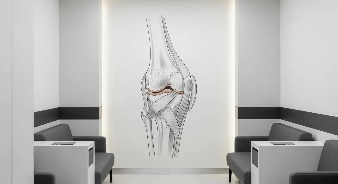
The teardrop muscle is a vital component of the thigh, known anatomically as the vastus medialis. It plays a crucial role in the functionality of the knee and contributes to overall stability.

Anatomy of Vastus Medialis
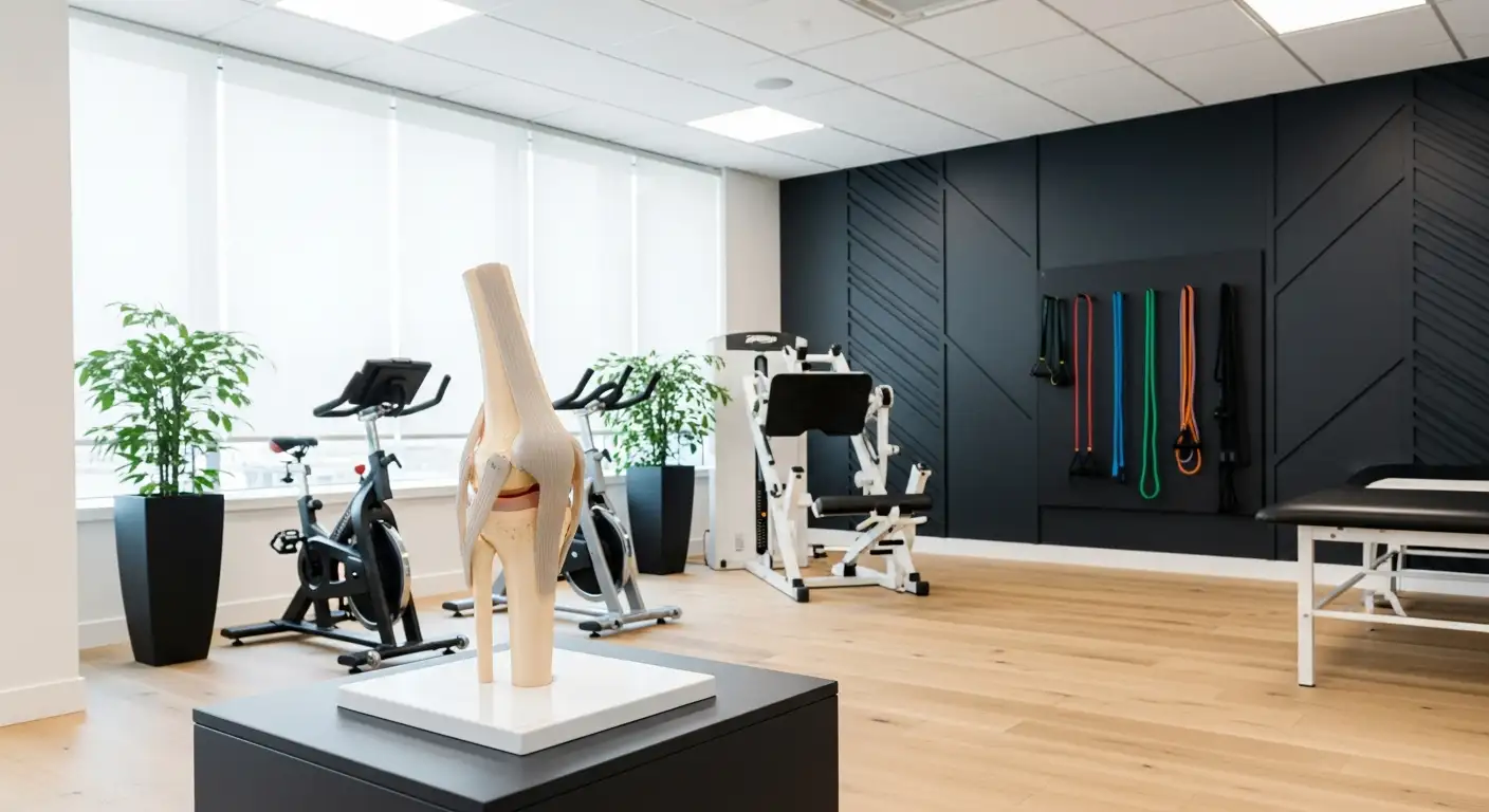
The vastus medialis, or teardrop muscle, is one of four muscles that make up the quadriceps group. It is located on the upper inside of the knee, extending down to the knee joint. This muscle is pivotal for extending the leg at the knee and stabilizing the patella, commonly referred to as the kneecap [1].
MuscleLocationFunctionVastus Medialis (Teardrop)Upper inner thighExtends the knee, stabilizes the patella
Weakness or injury in the vastus medialis can lead to issues with mobility, especially during weight-bearing activities such as running or climbing stairs [2].
Role in Knee Stability
The vastus medialis plays a significant role in knee stability. By ensuring the patella remains properly aligned during movements, it helps prevent knee injuries and promotes efficient movement patterns. The muscle contracts to help control patellar tracking, which is essential for maintaining knee health.
When the vastus medialis is weak or inactive, it can lead to improper tracking of the kneecap, potentially resulting in conditions such as patellofemoral pain syndrome. This underscores the importance of properly developing and maintaining strength in the teardrop muscle for overall knee health.
For further information on how to strengthen the vastus medialis, consider exploring targeted exercises or rehabilitation strategies listed in related sections of this article, including knee wraps for pain and osgood schlatter stretches.
Importance of VMO Training
The vastus medialis obliquus (VMO), often referred to as the teardrop muscle, is integral to knee stability and overall lower body function. Proper training of this muscle can yield significant benefits in preventing injuries and ensuring proper patella tracking.
Preventing Knee Injuries
Specific training of the VMO is crucial for maintaining patella position and limiting knee injuries. Weakness or fatigue in this muscle can lead to incorrect tracking of the patella during movement, which puts extra stress on the knee joint. This misalignment can result in various injuries such as patellofemoral pain syndrome, anterior cruciate ligament (ACL) tears, and tendinitis [1].
By incorporating exercises that strengthen the VMO, individuals can enhance their knee stability, reduce the risk of injuries during physical activities, and improve overall athletic performance. A well-conditioned VMO can help absorb shock and minimize strain on surrounding ligaments and cartilage.
Injury TypeCausePatellofemoral Pain SyndromeWeakness or fatigue in VMO leading to mal-trackingACL RuptureInadequate support from muscles around the jointChondromalaciaExcessive pressure on the knee jointTendinitisOveruse and improper tracking of the patella
Patella Tracking
The VMO plays a critical role in the tracking of the patella, or kneecap, during knee movement. Proper functioning of the VMO ensures that the patella moves fluidly within its groove on the femur. If the VMO is weak, the patella may shift away from its normal path, leading to discomfort and potential injury [2].
Strengthening the VMO can help correct mal-tracking issues. When the VMO is activated correctly, it pulls the patella medially (toward the center of the body) and stabilizes its movement. This alignment is particularly important during activities that involve bending and straightening of the knee, such as squatting, running, and climbing stairs.
For those who experience issues like a stabbing pain in the knee cap or wish to improve their knee stability, focusing on the VMO and implementing targeted exercises can prove highly beneficial. Strengthening the teardrop muscle not only enhances aesthetic appeal but also promotes better performance in athletic endeavors and everyday activities.
VMO Weakness and Knee Pain
The vastus medialis obliquus (VMO) muscle plays a critical role in knee health. Weakness in this muscle is closely linked to various knee pain conditions, most notably patellofemoral pain syndrome. This syndrome involves pain around the kneecap and can severely impact mobility and quality of life.
Link to Patellofemoral Pain Syndrome
Patellofemoral pain syndrome (PFPS) is commonly associated with weakness in the VMO. This weakness leads to mal-tracking of the patella, which can increase stress on the knee joint. Research indicates that the specific training of the VMO is crucial for maintaining proper patellar positioning and limiting knee injuries. Misfiring or fatigue of the VMO can exacerbate issues by causing the patella to track incorrectly, contributing to conditions such as chondromalacia and anterior cruciate ligament (ACL) injuries.
The following table summarizes the potential injuries related to VMO weakness:
ConditionDescriptionPatellofemoral Pain SyndromePain around the kneecap due to mal-trackingACL RuptureTear of the anterior cruciate ligament, often resulting from instabilityChondromalaciaSoftening and degeneration of the cartilage beneath the kneecapTendinitisInflammation of the tendons around the knee, which can flare due to improper tracking
Potential Injuries
Inadequate strength in the VMO can lead to a higher risk of knee instability and various injuries. Injuries may manifest as difficulty in walking, navigating stairs, or performing weight-bearing activities. Running and jumping can particularly strain the VMO, increasing the likelihood of strains or tears, as well as potential nerve and connective tissue damage.
Recognizing the importance of the VMO in knee health is essential for understanding knee pain. Proper training and rehabilitation strategies can help address VMO weaknesses, thereby reducing the risk of patellofemoral pain syndrome and other associated injuries. For insights into managing knee pain effectively, you can explore our articles on kneeling down and knee wraps for pain.
Assessing VMO Function
Effectively evaluating the vastus medialis obliquus (VMO) is important for understanding its functionality and performance in knee stability. Various methods can be employed to assess the VMO, including electromyography and targeted rehabilitation strategies.
Electromyography Evaluation
Electromyography (EMG) is a valuable diagnostic tool that can be used to assess the electrical activity of the VMO muscle. This technique helps in detecting abnormalities, weakness, or fatigue, which can be crucial for establishing appropriate rehabilitative plans aimed at correcting issues and preventing injuries [1].
EMG works by placing electrodes on the skin surface over the muscle of interest, allowing for the recording of electrical impulses during muscle contraction. The data collected can provide insights into the biomechanics of knee movement and highlight any dysfunction in the VMO.
EMG MeasurementSignificanceElectrical ActivityIndicates muscle activation and strengthFatigue LevelHelps identify if the muscle is overworked or weakResponse to StrengtheningAssesses improvements in rehabilitation
This information is essential for tailoring rehabilitation programs that specifically target the VMO to improve its performance.
Rehabilitation Strategies
Once a VMO injury or weakness has been identified, effective rehabilitation strategies can be implemented. Recovery typically involves physical therapy that focuses on exercises that strengthen the hip, thigh, and knee Verywell Health. The following exercises are commonly recommended:
ExercisePurposeStatic Quad StretchImproves flexibilityHip Flexor StretchIncreases mobility in the hip areaStraight Leg RaisesBuilds strength in the quadricepsStep-UpsEnhances functional strength and coordinationWall SquatsStrengthens the VMO while maintaining knee stability
Recovery from a VMO-related injury can take approximately six to eight weeks, depending on the severity. Severe cases may require surgical intervention, with post-operative recovery lasting longer. Continuous evaluation through methods like EMG can help monitor progress throughout rehabilitation.
For more detailed guidance on knee management and injury prevention, consider exploring related topics such as knee wraps for pain or genu recurvatum syndrome.
Developing Teardrop Quads
Focusing on the development of the teardrop muscle, which is part of the vastus medialis, involves targeted exercises and muscle isolation techniques. These methods help in enhancing the shape and strength of the quadriceps, contributing to knee stability and overall lower body aesthetics.
Targeted Exercises
To build the teardrop muscle effectively, several exercises specifically target the vastus medialis. The following table outlines various recommended exercises along with their respective benefits:
ExerciseBenefitsWide-Stance Hack SquatsEmphasizes the inner thigh and vastus medialis.Single-Leg ExtensionImproves stability and strengthens the teardrop muscle.Single-Leg Smith LungeEnhances balance and coordination in the lower body.Close-Foot Leg PressSpecifically targets the inner part of the quadriceps.Close-Foot Smith SquatsFocuses on developing the teardrop shape in the quads.
Incorporating single-leg movements, such as the single-leg press and extension, aids in conditioning and promoting muscle growth. Exercises like these improve balance, coordination, and symmetry among the leg muscles [3]. Understanding the anatomy of the quadriceps, including the rectus femoris and vastus lateralis, is crucial for targeting these exercises effectively [4].
Muscle Isolation Techniques
Isolating the teardrop muscle requires concentration on the muscle during workouts. Specific foot positions and techniques can enhance muscle engagement. Key tips include:
These techniques help ensure that workouts effectively target the teardrop muscle while minimizing involvement from surrounding muscle groups. For optimal results, individuals should incorporate these strategies into their workout regime regularly. For further assistance with knee pain or rehabilitation, consider exploring knee wraps for pain or seeking advice from professionals experienced in normal knee function and stabbing pain in knee cap.
Managing Quadriceps Strains
Quadriceps strains are common injuries, especially among athletes and individuals engaging in high-intensity sports. Understanding the causes of these muscle injuries and exploring effective treatment and rehabilitation strategies is critical for recovery.
Causes of Muscle Injuries
Quadriceps strains generally occur when the muscle is stretched beyond its limit, leading to tears in the muscle fibers near the tendon attachment. This can result in significant pain and impaired function. Key mechanisms that cause quadriceps strains include:
The rectus femoris, one of the quadriceps muscles, is frequently injured due to its dual role in both hip flexion and knee extension, among other predisposing factors such as muscle architecture and fiber type distribution. For a detailed overview of muscle strains, refer to OrthoInfo - AAOS.
Severity GradeDescriptionHealing TimeGrade 1Mild strain with limited painDays to weeksGrade 2Moderate strain with more significant pain and swellingWeeks to monthsGrade 3Severe tear with substantial loss of functionMonths
Treatment and Rehabilitation
Effective management of quadriceps strains often involves following the RICE protocol, which includes:
In addition, physical therapy plays an essential role in rehabilitation. Physical therapy focuses on improving range of motion and strength while ensuring the muscle is completely healed before returning to athletic activities to prevent reinjury. Techniques may also involve knee mobilization and targeted excercises for the quadriceps muscle.
For thorough assessment and rehabilitation strategies, consulting resources such as Physio-Pedia can be beneficial. In cases of persistent pain or complex injuries, seeking a professional evaluation may be necessary.
Properly managing quadriceps strains ensures that individuals can effectively return to their activities while minimizing the risk of future injuries.
References
[2]:
[3]:
[4]: